Home » Without Label » Abdominal Anatomy - Abdominal Anatomy Medical Illustration Medivisuals : The component of the urinary system, kidney and the ureter.
Abdominal Anatomy - Abdominal Anatomy Medical Illustration Medivisuals : The component of the urinary system, kidney and the ureter.
Abdominal Anatomy - Abdominal Anatomy Medical Illustration Medivisuals : The component of the urinary system, kidney and the ureter.. It follows the thorax or cephalothorax. We'll identify as many organs as we can, see how they fit into the. The anterior abdominal wall extends from the xiphoid and lower six costal cartilages to the anterior aspect of the pelvic bones. Then liver & spleen) palpate 4 quadrants abdomen (superficial then deep) assess for kidney area pain (cvat) wash hands time target: The abdomen is the part of the body that contains all of the structures between the thorax (chest) and the pelvis, and is separated from the thorax via the diaphragm.
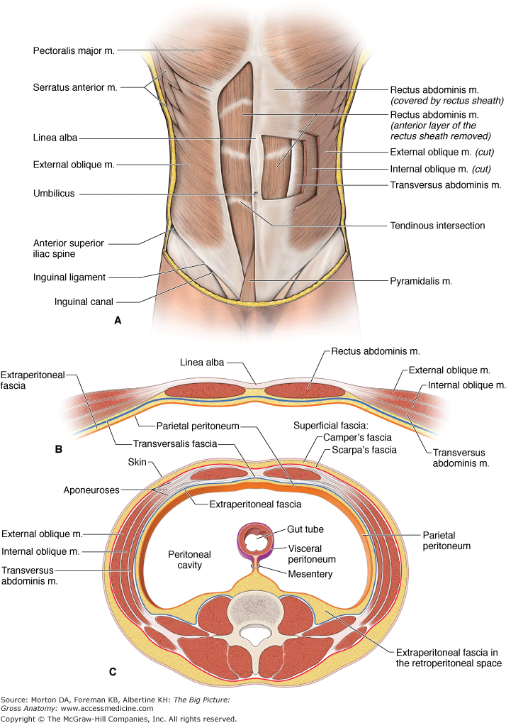
Overview the abdomen contains many vital organs: Skin, superficial fascia, muscles and associated fascia, and parietal peritoneum. The anterior abdominal wall extends from the xiphoid and lower six costal cartilages to the anterior aspect of the pelvic bones. Together, these three turn nutrients into usable energy, as well as help dispose of solid waste. In anatomy and physiology, you'll learn how to divide the abdomen into nine different regions and four different quadrants.
Abdominal Aorta | Geeky Medics from geekymedics.com Abdominal anatomy includes a major element of the gastrointestinal, system, the caudal end of the oesophagus, stomach, large and small intestine, liver, pancreas and the gallbladder. It is the long, flat muscle that extends vertically between the pubis and the fifth, sixth, and seventh ribs. It is bounded superiorly by the xiphoid process and costal margins, posteriorly by the vertebral column and inferiorly by the pelvic bones and inguinal ligament. Together, these three turn nutrients into usable energy, as well as help dispose of solid waste. Connective tissue called the mesentery holds the abdominal organs together. The rectus abdominis connects to the xiphoid process, a bony landmark at the bottom of the sternum. Inferiorly the abdomen is open to the pelvis, communicating through the superior pelvic aperture (pelvic inlet). The abdominal aorta enters the abdomen through the diaphragm at the level of the twelfth thoracic vertebre and continues to just below the umbilical area, where it splits into the right and left common iliac arteries.
It is the long, flat muscle that extends vertically between the pubis and the fifth, sixth, and seventh ribs.
The rectus abdominis connects to the xiphoid process, a bony landmark at the bottom of the sternum. The aorta is the largest blood vessel in the body. If you plan to enter a healthcare profession such as nursing, this is something you'll use on the job when performing abdominal assessments (and while documenting). These two apertures, together with abdominal walls, bound the abdominal cavity. Observe abdomen (shape, contours, scars, color, etc) auscultate abdomen (bowel sounds, bruits) percuss abdomen (general; Connective tissue called the mesentery holds the abdominal organs together. The abdomen is the part of the body that contains all of the structures between the thorax (chest) and the pelvis, and is separated from the thorax via the diaphragm. The abdominal cavity is the part of the body that houses the stomach, liver, pancreas, kidneys, gallbladder, spleen, and the large and small intestines.the diaphragm marks the top of the abdomen and the horizontal line at the level of the top of the pelvis marks the bottom. Inferiorly the abdomen is open to the pelvis, communicating through the superior pelvic aperture (pelvic inlet). It is the most complete reference of human anatomy available on web, ipad, iphone and android devices. This mri abdomen axial cross sectional anatomy tool is absolutely free to use. Skin, superficial fascia, muscles and associated fascia, and parietal peritoneum. The major organs of the abdomen include the small intestine, large intestine, and stomach.
The major organs of the abdomen include the small intestine, large intestine, and stomach. It is bounded superiorly by the xiphoid process and costal margins, posteriorly by the vertebral column and inferiorly by the pelvic bones and inguinal ligament. These two apertures, together with abdominal walls, bound the abdominal cavity. The image also shows the pelvis, uterus, and urinary. The abdominal cavity is the part of the body that houses the stomach, liver, pancreas, kidneys, gallbladder, spleen, and the large and small intestines.the diaphragm marks the top of the abdomen and the horizontal line at the level of the top of the pelvis marks the bottom.
Abdominal anatomy, illustration - Stock Image - F029/5259 ... from media.sciencephoto.com The region occupied by the abdomen is called the abdominal cavity, and is enclosed by the abdominal muscles at front and to the sides, and by part of the vertebral column at the back. The rectus abdominis connects to the xiphoid process, a bony landmark at the bottom of the sternum. The stomach, the small intestine (jejunum and ileum), the large intestine (colon), the liver, the spleen, the gallbladder, the pancreas, the uterus, the fallopian tubes, the ovaries, the kidneys, the ureters, the bladder, and many blood vessels (arteries and veins). Then liver & spleen) palpate 4 quadrants abdomen (superficial then deep) assess for kidney area pain (cvat) wash hands time target: Stomach is a muscular bag forming the most distensible part of the human digestive system. The majority of these organs are encased in a protective membrane termed the peritoneum. Use the mouse scroll wheel to move the images up and down alternatively use the tiny arrows (>>) on both side of the image to move the images.>>) on both side of the image to move the images. The abdominal aorta enters the abdomen through the diaphragm at the level of the twelfth thoracic vertebre and continues to just below the umbilical area, where it splits into the right and left common iliac arteries.
The anterior abdominal wall extends from the xiphoid and lower six costal cartilages to the anterior aspect of the pelvic bones.
The aorta is the largest blood vessel in the body. Connective tissue called the mesentery holds the abdominal organs together. Observe abdomen (shape, contours, scars, color, etc) auscultate abdomen (bowel sounds, bruits) percuss abdomen (general; Together, these three turn nutrients into usable energy, as well as help dispose of solid waste. The abdominal wall surrounds the abdominal cavity, providing it with flexible coverage and protecting the internal organs from damage. Inferiorly the abdomen is open to the pelvis, communicating through the superior pelvic aperture (pelvic inlet). We're going to take apart a plastic anatomy model and see what we can find in the abdomen. The majority of these organs are encased in a protective membrane termed the peritoneum. We'll identify as many organs as we can, see how they fit into the. The anterolateral abdominal wall consists of four main layers (external to internal): Abdomen, in human anatomy, the body cavity lying between the chest or thorax above and the pelvis below and from the spine in the back to the wall of abdominal muscles in the front. The diaphragm is its upper boundary. The abdominal aorta enters the abdomen through the diaphragm at the level of the twelfth thoracic vertebre and continues to just below the umbilical area, where it splits into the right and left common iliac arteries.
If you plan to enter a healthcare profession such as nursing, this is something you'll use on the job when performing abdominal assessments (and while documenting). The stomach, the small intestine (jejunum and ileum), the large intestine (colon), the liver, the spleen, the gallbladder, the pancreas, the uterus, the fallopian tubes, the ovaries, the kidneys, the ureters, the bladder, and many blood vessels (arteries and veins). The aorta is the largest blood vessel in the body. Together, these three turn nutrients into usable energy, as well as help dispose of solid waste. The majority of these organs are encased in a protective membrane termed the peritoneum.
Abdominal Anatomy / Process Of Digestion - How The ... from accessmedicine.mhmedical.com The abdominal wall surrounds the abdominal cavity, providing it with flexible coverage and protecting the internal organs from damage. The major organs of the abdomen include the small intestine, large intestine, and stomach. It follows the thorax or cephalothorax. The image also shows the pelvis, uterus, and urinary. The diaphragm is its upper boundary. Ct, mri, radiographs, anatomic diagrams and nuclear images. Abdomen, in human anatomy, the body cavity lying between the chest or thorax above and the pelvis below and from the spine in the back to the wall of abdominal muscles in the front. Abdomen anatomy the abdomen is comprised primarily of the digestive tract and other accessory organs which assist in digestion, the urinary system, spleen, and the abdominal muscles (shown below).
The rectus abdominis connects to the xiphoid process, a bony landmark at the bottom of the sternum.
It also contains the spleen. It is composed of several layers, including skin, superficial fascia, subcutaneous fat, anterolateral and midline muscle groups, transversalis fascia, extraperitoneal fat and peritoneum. The region occupied by the abdomen is called the abdominal cavity, and is enclosed by the abdominal muscles at front and to the sides, and by part of the vertebral column at the back. Abdomen, in human anatomy, the body cavity lying between the chest or thorax above and the pelvis below and from the spine in the back to the wall of abdominal muscles in the front. The liver, stomach, and abdominal contents are clearly identified and labeled, including the cecum, ascending colon, transverse colon, descending colon, and small intestine. The abdomen (colloquially called the belly, tummy, midriff or stomach) is the part of the body between the thorax (chest) and pelvis, in humans and in other vertebrates.the abdomen is the front part of the abdominal segment of the trunk.the area occupied by the abdomen is called the abdominal cavity.in arthropods it is the posterior tagma of the body; Overview the abdomen contains many vital organs: Then liver & spleen) palpate 4 quadrants abdomen (superficial then deep) assess for kidney area pain (cvat) wash hands time target: The major organs of the abdomen include the small intestine, large intestine, and stomach. Abdominal anatomy includes a major element of the gastrointestinal, system, the caudal end of the oesophagus, stomach, large and small intestine, liver, pancreas and the gallbladder. Use the mouse scroll wheel to move the images up and down alternatively use the tiny arrows (>>) on both side of the image to move the images.>>) on both side of the image to move the images. These organs are held together loosely by connecting tissues. Together, these three turn nutrients into usable energy, as well as help dispose of solid waste.